Pediatric basic life support (PBLS) is a major component of the emergency medical response to the pediatric victims with cardiac arrest, which should be adequately implemented for a series of survival processes. The pediatric chain of survival comprises five components, including prevention and early recognition of cardiac arrest, early access (activation of emergency medical system [EMS]), early high-quality cardiopulmonary resuscitation (CPR), early defibrillation, and effective advanced life support and post-cardiac arrest care (Fig. 1). The first four processes of the survival chain correspond to PBLS. Similar to adults, in children, prompt and effective CPR is essential for the successful recovery of spontaneous circulation and good neurological outcomes. In children, the survival rate varies depending on the cause of the cardiac arrest. For a cardiac arrest due to an asphyxial arrest, the rate of neurologically normal survival is 70%, while the survival rate is 20% to 30% in the case of a cardiac arrest from ventricular fibrillation (VF) [1]. Neonate is defined as being hospital after birth. An infant is defined as being less than 1 year of age after neonatal period. PBLS guideline applies to children under 8 years of age except neonate for both of the lay rescuer and the health care provider. As cardiac arrest from asphyxial arrest is a lot more common in children compared to those due to VF, ventilation is extremely important in pediatric CPR.
PBLS FOR LAY RESCUERS
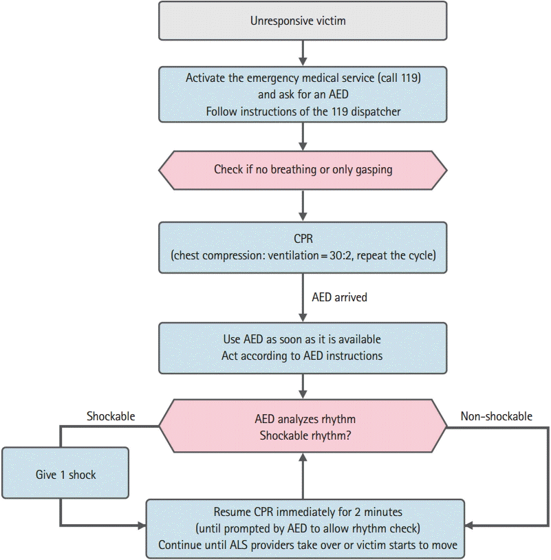
The algorithm for CPR in infants and pediatric patients experiencing cardiac arrest is the same as that used in adults, signifying unification in the education and training as well as continuation of the existing the 2011 Korean CPR guidelines, according to which chest compressions are carried out followed by rescue breathing [2-4]. A flow diagram of PBLS for lay rescuers is shown in Fig. 2.
1. Safety of the rescuer and the patient
When conducting CPR, the safety of the rescuer and patient must be confirmed at the current location. CPR may carry the risk of transmission of infectious diseases, but the actual risk to the rescuer is extremely low.
2. Determination of the unresponsiveness
If an unresponsive patient is gasping or not breathing at all, the lay rescuer should understand that the patient is undergoing a cardiac arrest and requires CPR. The rescuer should lightly tap the patient and shout out ŌĆ£are you okay?ŌĆØ or call out the name of the patient if known. Then, the rescuer should promptly check whether the child has injuries or requires medical treatments.
3. Activation of the EMS
If the patient shows no response to stimulation and if the rescuer is the only person present, the rescuer should shout for help. If someone else arrives for help, the rescuer should request him or her to make a call to 119 as well as find an automated external defibrillator (AED). If there is no one else nearby, the rescuer should make a rescue request to 119 promptly. If the rescuer has a personal cellular phone, the rescuer should promptly call 119 at the site itself, without leaving the child. The rescuer should let the 119 dispatcher know about the status of the unresponsive patient, request for an AED, and perform the next resuscitation steps by following instructions given by the dispatcher.
If the patient shows no response to stimulation and if there are two or more witnesses, the first rescuer should start CPR immediately and the other should call an EMS and request for an AED.
Most cases of cardiac arrests in children are due to asphyxial arrest rather than VF. Therefore, if there is only one rescuer without a cellular phone, the rescuer should perform CPR for the first 2 minutes then call an EMS and bring a nearby AED. The rescuer should return to the patient as soon as possible and use the AED. If no AED can be found, the rescuer should resume CPR starting with chest compression.
4. Checking patient breathing
If a patient is confirmed to be breathing regularly, the child is not in need of CPR. In such cases, after confirming that there is no evidence of physical injuries, the rescuer should turn the child onto one side and into the recovery position to keep their airway open and reduce the risk of aspiration. The breathing status of the patient should be checked continuously until the arrival of the EMS. If the child tries to change his or her position into a more relaxed position, the child should be allowed to do so.
If a patient is not responding to stimulation and is not breathing or only gasping (cardiac arrest breathing), CPR should be started. In some cases, patients who are in need of CPR and gasping can be mistaken as having normal breathing. Patients who are gasping should be regarded as not breathing, for which CPR should be started.
5. Chest compression
In CPR, adequate chest compression maintains blood flow to vital organs and increases the possibility of recovery of spontaneous circulation. If a child is neither responding to stimulation nor breathing, the rescuer should immediately perform chest compression 30 times. Chest compression should be performed at a speed of 100 to 120 per minute and by pressing down at least one-third the depth of the anteroposterior diameter of the rib cage (chest thickness) or 4 cm in infants and 4 to 5 cm in children [5-7]. It is best to perform chest compression by laying the patient on a flat and hard surface.
For infants, the lay rescuer or a healthcare provider who is performing the CPR should use two fingers to compress the center of the chest just below the line between the nipples. For children, the rescuer should compress the lower half of the sternum with one or both hands. Special attention should be paid to avoid pressing of the xiphoid process or the rib cage. Proper depth should be maintained for each compression, irrespective of whether one or both hands are used, and the chest must be allowed to recoil to its normal position after every compression.
After each compression, the chest must be allowed to recoil fully, as proper venous return to the heart can be completed only when the chest is fully relaxed. During pediatric CPR, incomplete chest recoil is common, particularly when rescuers are fatigued. Incomplete chest relaxation increases pressure inside the chest cavity and reduces venous return, coronary perfusion, cardiac output, and cerebral perfusion [8,9].
Fatigue of the rescuer can lead to inadequate speed and depth of chest compression, as well as chest recoil. Even if the rescuer denies fatigue and continues CPR, the quality of chest compression will decrease within a few minutes [10,11]. If there are two or more rescuers, they should take turns to perform CPR every 2 minutes in order to prevent fatigue and reduction in the quality and speed of chest compression. The switching between the rescuers to perform chest compression should be done as soon as possible (ideally within 5 seconds) to minimize the interruption in chest compression. Each rescuer should perform 30 chest compressions and 2 ventilations, until an EMS arrives or the patient starts breathing. For CPR in infants and children, chest compression and ventilation must be provided together to yield the best outcome. CPR accompanied by both ventilation and chest compressions must be performed by a person trained in performing infant or pediatric CPR, both inside and outside the hospital. However, if a rescuer is not trained in artificial ventilation or is unable to perform it, he or she should perform chest compression until an EMS arrives [12,13].
6. Opening the airway and giving ventilation
As infants or children who are not responding may have their airway obstructed by their tongues, their airway must be opened by tilting the head and lifting the chin for both injured and noninjured patients. For infants, mouth-to-mouth-and-nose technique should be performed. Mouth-to-mouth should be used for children. While blowing the breath in, the rising of the chest should be confirmed and each breath should take about 1 second. If the chest does not rise, the head should be tilted and the mouth should be adequately covered to prevent leaking of the air before trying ventilation again. While performing ventilation for infants, if it is difficult to cover both the mouth and nose at once, mouth-to-mouth or mouth-to-nose breathing may be performed. For mouth-to-mouth, the nose should be occluded, and for mouth-to-nose, the mouth should be occluded.
7. Ratio of chest compression to ventilation
After performing 30 compressions, two ventilations should be performed immediately. If there is only one rescuer, 2 ventilations following 30 chest compressions should be performed, with the shortest possible break in order to minimize the duration of interruption of chest compression. When there are two rescuers, one should be in charge of chest compression and the other in charge of ventilation to sequentially perform 30 chest compressions and 2 ventilations. Chest compression and ventilation must not be performed simultaneously. Interruption of chest compression should be minimized. It would take approximately 2 minutes for one rescuer to perform 5 cycles of chest compression and ventilation at the ratio of 30:2. Two rescuers should perform chest compression and ventilation by rotating after every 5 cycles (Table 1).
PBLS FOR HEALTHCARE PROVIDERS
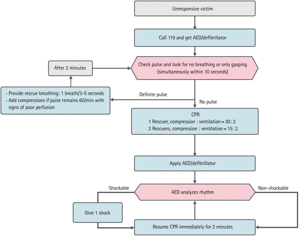
PBLS for healthcare provider is essentially very similar to PBLS for the lay rescuer, except a few differences [14,15]. As healthcare providers usually work in teams rather than individually and because a series of activities is performed concurrently (for example, chest compression and preparation for ventilation), the priority of each activity is relatively less emphasized. A flow diagram of PBLS for healthcare providers is shown in Fig. 3.
1. Checking for patient response and breathing
If an unresponsive patient is gasping or not breathing at all, the rescuer should understand that the patient is undergoing a cardiac arrest and requires CPR. The rescuer should lightly tap the patient and shout out ŌĆ£are you okay?ŌĆØ or call out the name of the patient if known. Then, the rescuer should promptly check whether the child has injuries or requires medical treatments.
2. Activation of the EMS
If a patient is neither responding nor breathing (or he/she has abnormal breathing such as gasping), the rescuer should ask nearby individuals to activate an EMS and request for an AED.
3. Checking patient pulse
If an infant or a child is neither responding nor breathing normally, a healthcare provider should check for their pulse within 10 seconds. For infants, the pulse should be checked on the brachial artery, and for children, on the carotid or femoral artery. If a pulse is not detected within 10 seconds or if it is uncertain, chest compression should be started [2].
1) Case of palpable pulse and inadequate breathing
If a pulse is detected to be more than 60 per minute but breathing is inadequate, ventilation should be provided at the speed of 12 to 20 times per minute (1 breath every 3 to 5 seconds) until the return of spontaneous breathing. The pulse should be rechecked every 2 minutes, and each pulse check should not exceed 10 seconds.
2) Case of bradycardia and poor systemic perfusion
If the pulse rate is less than 60 per minute and the perfusion status is poor (i.e., pale skin, patches of spots, or cyanosis is observed) even with oxygen and adequate ventilation, chest compression should be performed. As the cardiac output of infants and children depends greatly on the heart rate, bradycardia with poor systemic perfusion signals the necessity for chest compression. Performing CPR immediately after the occurrence of cardiac arrest can lead to an increased survival rate. Although the critical value of the heart rate at which chest compression should be performed is not yet clearly defined, chest compression is recommended for a heart rate less than 60 per minute and poor perfusion for the convenience of education and ease of memorization.
4. Chest compression
Chest compression should be performed if infants or children do not respond, are not breathing, and do not have a pulse (or if it is unclear whether they have a pulse). The differences between a healthcare provider and a lay rescuer are in the method of chest compression in infants. When a healthcare provider is alone, chest compression using two fingers is performed for an infant. The two-thumb encircling method for chest compression is performed when there are two or more rescuers. The sides of the thorax of an infant are wrapped around with open hands, with the two thumbs side-by-side on the sternum for strong compression. The advantage of the two-thumb encircling method is that it increases perfusion pressure of the coronary artery to a greater extent compared to the two-finger chest compression, allowing pressure depth and strength consistency to generate higher systolic and diastolic blood pressures [16-18]. If it is not possible to wrap the side of the thorax with both hands, the chest should be compressed using two fingers. The location for chest compression should be the lower half of the sternum in children and the sternum just below the nipple line in infants.
5. Opening the airway and giving ventilation
After 30 chest compressions (15 compressions in the case of two rescuers), the airway is opened using head-tilt chin-lift maneuver followed by 2 ventilations. If there are signs of spinal cord injury, head tilting should not be performed. Instead, the airway should be opened using the jaw-thrust method. As proper ventilation with an opened airway is extremely important for pediatric CPR, head-tilt chin-lift maneuver should be performed if jaw-thrust is not sufficient to open the airway.
6. Ventilation
1) Method of ventilation
If it is difficult to concurrently cover the mouth and nose while performing ventilation on infants, mouth-to-mouth or mouth-to-nose breathing may be performed. When mouth-to-mouth breathing is performed, the nose should be closed. For mouth-to-nose breathing, the mouth should be closed. In both cases, rising of the chest should be confirmed during ventilation.
2) Ventilation method to prevent hyperventilation
Before establishing an advanced airway (such as endotracheal intubation, or supraglottic airway), ventilation is performed twice after 30 chest compressions (one rescuer) or 15 chest compressions (two healthcare provider rescuers), with mouth-to-mouth breathing or the bag-mask method. After establishing an advanced airway, the ŌĆ£compression-ventilation ratioŌĆØ of CPR is not used. Chest compression at the speed of 100 to 120 per minute and 10 ventilations per minute are performed consistently. Two or more healthcare providers take turns in performing compression every 2 minutes to prevent the rescuers from getting fatigued. If the perfusion rhythm returns but there is no breathing, ventilation is performed 12 to 20 times per minute (1 ventilation every 3 to 5 seconds). Excessive ventilation during CPR reduced venous return, leading to decreased cardiac output and cerebral blood flow, and increases pressure within the thoracic cavity, leading to decreased coronary artery perfusion [19]. Therefore, rescuers should perform ventilation by following the recommended frequency of ventilation. Because manual bag-mask ventilation can provide high pressure, ventilation is performed to the extent in which the chest rise is just observed.
3) Two-rescuer bag-mask ventilation
In the case with the difficulty in attaching the mask, severe obstruction in the airway, or poor lung elasticity, bag-mask ventilation performed by two rescuers together can be useful and helpful in providing effective bag-mask ventilation to the patients. One rescuer should maintain the airway and tightly seal the mask onto the face using both the hands while the other compresses the ventilation bag. Both rescuers should confirm rise of the chest.
4) Gastric inflation and cricoid pressure
As gastric inflation may prevent effective ventilation and induce regurgitation, it should be avoided. To minimize gastric inflation, each ventilation should be performed for 1 second to prevent excessive pressure during exhalation. In addition, the application of cricoid pressure can be considered although its application is not recommended in a routine procedure. It should be considered only when the patient is unconscious and there is another healthcare provider to provide help, and it should be performed with caution as excessive cricoid pressure can block the airway.
5) Oxygen
One hundred percent oxygen is provided during CPR, because there has been no report of any harmful effects of differing oxygen concentrations in humans, after the neonatal period. When the patientŌĆÖs condition has stabilized, oxygen should be provided with monitoring of the saturation. The provision of humidified oxygen can prevent the drying of the mucous membrane and thickening of lung secretions. Oxygen is provided using a mask or a nasal cannula.
7. Ratio of chest compression and ventilation
One rescuer performs chest compression and ventilation at the ratio of 30:2. When two rescuers perform CPR on infants or children, one performs chest compression and the other performs ventilation at the ratio of 15:2, with opening of the airway. The interruption of chest compression for the ventilation should be minimized. After intubation, the ratio of chest compression and ventilation is no longer followed. Instead, the rescuer responsible for chest compression does not stop the compression for ventilation and performs it continuously at the speed of 100 to 120 per minute. The rescuer responsible for ventilation provides ventilation at the speed of 10 times per minute (1 ventilation every 6 seconds).
8. Defibrillation
VF may be the cause of sudden cardiac arrest or it may occur during CPR. The sudden collapse of child in the presence of another person (for example, a child who collapsed while exercising) that may be caused by VF or pulseless ventricular tachycardia, immediate CPR and prompt defibrillation are necessary in such cases. As VF or pulseless ventricular tachycardia may respond to defibrillation, they are categorized as ŌĆ£shockable rhythmsŌĆØ.
In infants, it is better to use a manual defibrillator by well-trained healthcare provider to give a shock. For defibrillation, the first energy dose is at 2 to 4 J/kg and the second is 4 J/kg or higher; the shock should not exceed the maximum dose for adults [20,21]. If a manual defibrillator is not available, an AED with a pediatric energy attenuator should be used. However, if both a manual defibrillator and an AED with a pediatric energy attenuator are lacking, an AED for adults without an attenuator can be used for infants.
Rescuers should minimize the time between chest compression and defibrillation, and resume CPR by restarting chest compression immediately after the defibrillation. The use of an AED will ensure that rescuers reanalyze the rhythm every 2 minutes, and defibrillation should be performed immediately after chest compression (Table 2).
9. Compression-only CPR
In infants and children, performing both chest compression and ventilation is the best CPR methods. As the most common cause of cardiac arrest in infants and children is asphyxia arrest, ventilation is required as a part of effective CPR [22]. Therefore, infant and pediatric CPR accompanies resuscitation comprising ventilation and chest compression. However, if artificial ventilation is not possible or the rescuer is not known about how to perform ventilation, compression-only CPR must be performed on its own [12,13].
RESUSCITATION UNDER SPECIAL CONDITIONS
1. Ventilation through tracheostomy
Caretakers of children with tracheostomy (parents, school nurses, or home healthcare providers) should know how to maintain the airway, clear airway discharges, and perform CPR through the artificial airway. Ventilation should be performed through tracheostomy while confirming airway maintenance and rising of the chest. If ventilation through tracheostomy is not effective even after effective suctions, maintenance of the airway should be reconfirmed. In patients who received tracheostomy, mouth-to-tracheostoma breathing is performed. When the tracheostoma is already sealed, bag-mask ventilation is performed through the nose or mouth [23].
2. Trauma
Although the criteria for performing CPR in children with injuries are the same as those in children with other diseases, some aspects should be emphasized. Errors in the opening and maintaining the airway, as well as errors due to unawareness of internal bleeding, commonly occur during pediatric resuscitation. Following are some precautions that must be taken while performing CPR on pediatric patients with injuries [24].
A suction device should be used if there is a possibility of airway obstruction due to broken tooth pieces or blood. If there is external bleeding, external pressure should be applied to stop the bleeding. Considering the mechanism of injury, if there is a possibility of spinal cord injury, the movement of the cervical spine should be minimized and the head or neck should not be pulled or moved. The airway should be opened using jaw-thrust, not head-tilt. If jaw-thrust is not sufficient to maintain the airway, head-tilt chin-lift maneuver can be performed. If there are two rescuers, one opens the airway and the other prevents the movement of the cervical spine. As infants and children have relatively large heads, the best position to prevent bending of the cervical spine can be achieved by placing the back of the head on a recessed location or fixing the body in a slightly raised position [25]. Children with multi-organ injuries should be transferred to a trauma center with a pediatric specialist if possible. Thoracotomy may be considered for children with penetrative injuries [26].
3. Drowning
In the case of drowning, the duration of submersion in water is an important factor for the prediction of prognosis. The age, promptness of emergency treatment, form of water (e.g., sea water), water temperature, and the presence or absence of witnesses are not reliable prognosis factors [27]. As the survival rate is relatively high in the case of drowning in ice water even with long submersion durations, the rescue time should be extended in such case [28,29]. Drowned children must start receiving CPR immediately after being rescued from the water. Rescuers with special training may start ventilation in water. Because chest compression is not efficient in water, it is not usually performed [30]. As there is no evidence that water causes airway obstruction, no time is wasted to pump out water from the lungs of the rescued person [31]. CPR should be started by performing chest compression and the airway is opened, ventilation is performed twice. If the rescuer is alone, 5 cycles of chest compression and ventilation are performed before calling EMS and preparing an AED. If there are two rescuers, one continues CPR while the other activates EMS and prepares an AED [32].