The 2020 Korean pediatric cardiopulmonary resuscitation (CPR) guideline is the medical recommendation derived on the basis of scientific evidence for pediatric basic life support (PBLS). This is based on the scientific consensus and treatment recommendations provided by the International Liaison Committee on Resuscitation in 2020, which provides the CPR guideline. Studies published for PBLS were additionally considered [1]. The evidence of revised items that are of high clinical importance and require further consideration were reviewed in an acceptable adaptation or hybrid format and meta-analysis or scoping review was used.
MAJOR CHANGES IN 2020 PBLS GUIDELINE
Compared with the 2015 guideline [2], the changes in the 2020 PBLS guideline are as follows.
Endotracheal intubation or supraglottic airway (SGA) insertion versus bag mask ventilation (BMV)
The use of BMV is recommended rather than endotracheal intubation or SGA insertion for pediatric out-of-hospital cardiac arrest (OHCA) (Class IIb, Level C-LD). For pediatric in-hospital cardiac arrest, there is no comparable evidence for applying endotracheal intubation or SGA insertion and BMV to date.
Dispatcher-assisted CPR
It is recommended to provide dispatcher-assisted CPR with the help of emergency services for pediatric OHCA (Class I, Level C-LD). If bystander CPR is not performed for OHCA, it is recommended to provide dispatcher-assisted CPR using the help of emergency services (Class I, Level C-LD). The recommendation to provide dispatcher-assisted CPR if the bystander CPR is performed for pediatric OHCA cannot be set with the evidence available to date.
Compression-only CPR
The rescuer should perform standard CPR with ventilation for infant and pediatric OHCA (Class I, Level B-NR). Although the bystander is unable to perform rescue breaths or has not been trained in it for pediatric OHCA, at least compression-only CPR should be done (Class I, Level B-NR).
SEQUENCE OF PBLS
Pediatric chain of survival
Basic life support (BLS) in pediatric cardiac arrest has a considerable impact on the return of spontaneous circulation (ROSC) and survival rate; it begins with the prevention of cardiac arrest, which is the first step in the chain of survival. The pediatric chain of survival begins with the recognition of cardiac arrest and a rescue request. However, before this, the prevention of cardiac arrest begins with several institutional strategies for injury prevention and safety outside the hospital. In the hospital, it is important to make efforts to prevent cardiac arrest using an early warning system. The first three among the five elements in the survival chain belong to BLS (Fig. 1). Similar to that in adults, for children, providing rapid and effective CPR by laypersons is helpful to regain successful ROSC and neurological recovery. For children, the survival rate considerably varies depending on age and cause of cardiac arrest, and it increases with age. For pediatric in-hospital cardiac arrest (IHCA), the survival rate is as high as >40% [3].
Importance of prevention in pediatric cardiac arrest
Cardiac arrest is difficult to predict and can occur in various places. Both non-medical factors such as cardiac arrest awareness and CPR education and medical factors such as prevention, treatment, and rehabilitation of cardiac arrest are all related to survival. The survival rate in cardiac arrest is low even with effective CPR; therefore, taking necessary precautions is important to prevent death from cardiac arrest. Respiratory failure and sudden infant death syndrome are the primary causes of infantile cardiac arrest. However, for children aged >1 year, trauma is the most common cause of cardiac arrest. Therefore, unlike that in adults, pediatric cardiac arrest can be prevented by appropriate management of the environment and changes in lifestyle. Sudden infant death syndrome can be prevented by not letting the child sleep on the stomach, not laying on a soft floor, and the caregiver’s smoking cessation. Traffic accidents, which are the primary cause of injury, can be prevented by wearing a seat belt and installing a child car seat. Child safety seats vary by age. Baby safety seats facing the rear should be used for infants aged <1 year and weighing <9 kg; child safety seats should be used for children aged 1 to 4 years, and a secondary chair with a seat belt is necessary for children aged 4 to 7 years. If children aged <12 years sit in the front seat, airbag-related fatal injuries can occur, and the risk from improper seat belting increases. Drowning is the second most frequent cause of accidental death in children aged <5 years as well as the third most frequent cause of accidental death in adolescents [2]. Most children drown in swimming pools while their caregivers are away. Adolescents often drown in either lake or river. Drowning can be prevented by wearing lifesaving gear during swimming. Recent domestic statistics show that murder by assault is the third cause of death among children aged between 1 and 9 years. Efforts to prevent such accidents should be made by attention and active reporting of child abuse. In adolescents aged >10 years, the first cause of death is deliberate self-harm (suicide). It can be prevented by providing emotional support to adolescents and active interventions against suicide risk [4].
Recognition of cardiac arrest, rescue request, and bystander CPR
Because an asphyxial cardiac arrest is more common in children, when a cardiac arrest is noticed, prompt CPR is as important as a rapid rescue request; however, because mobile phones are easily accessible in Korea, immediate rescue request should be made first, similar to that in adults. A domestic study on pediatric OHCA shows that the faster the CPR is performed after cardiac arrest, the higher the ROSC is, and rapid and effective layperson’s CPR at the scene improves the rate of ROSC in pediatric OHCA [5]. Furthermore, the neurological results are better when patients are discharged.
PBLS FOR LAY RESCUERS
There is a difference in the cause of cardiac arrest between children and adults. Moreover, because their sizes are different, CPR methods differ. However, it is difficult to distinguish between children and adults based on only one feature, and there is a lack of scientific evidence to determine the age at which different CPR methods should be applied. In this guideline, age was classified based on the applicability at the scene of cardiac arrest and educational ease. When a child is too big in size to be differentiated from an adult, it is the discretion of the rescuer to determine which method, pediatric or adult CPR, can be applied. The rescuer can apply either method because the miscalculation of the age of the patient does not cause significant harm to the cardiac arrest patient. The definition of age in CPR is listed as follows: neonate, up to 4 weeks from birth; infant, <1 year old; pediatrics, from 1 to <8 years old; and adult, ≥8 years old.
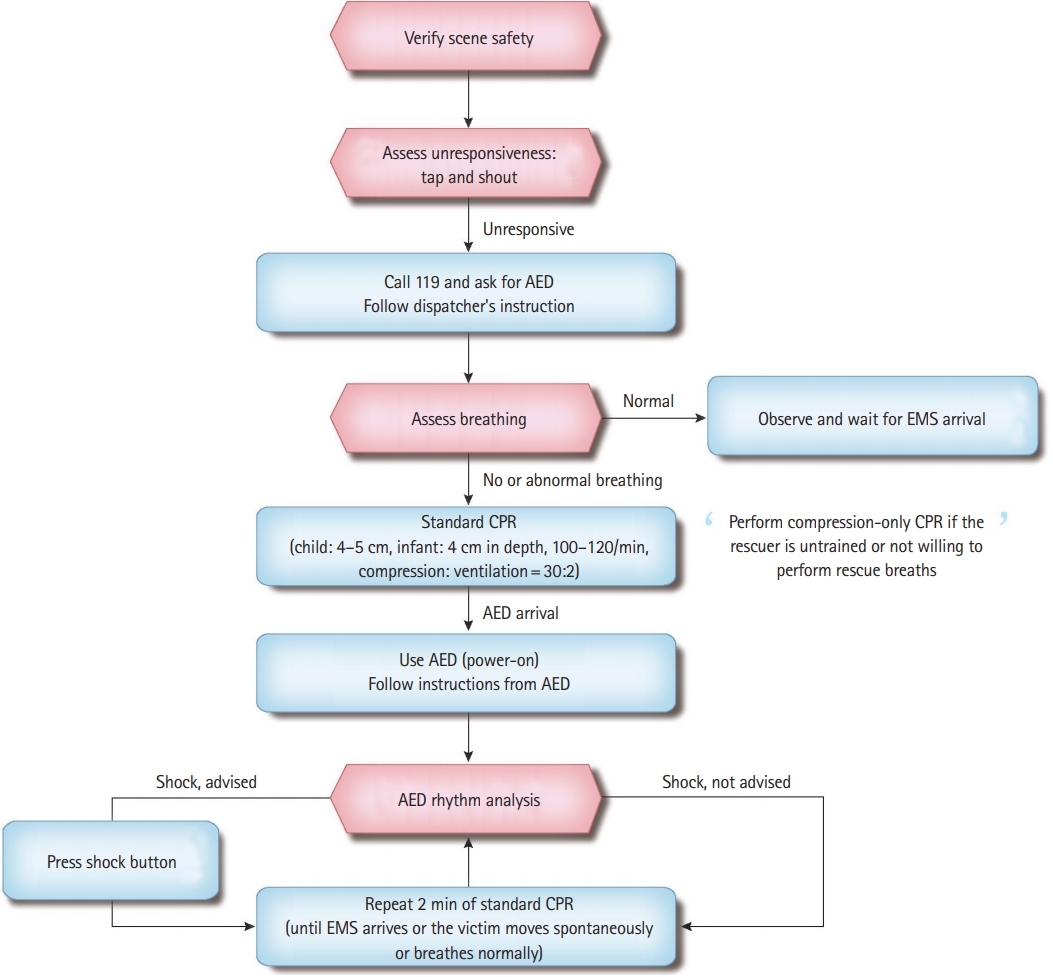
PBLS is applied to infants and children aged <8 years regardless of the layperson or the healthcare provider. Fig. 2 shows the algorithm for PBLS (Table 1). As shown in Fig. 3, for transmission risk of infectious diseases, such as severe acute respiratory syndrome and coronavirus disease-19 (COVID-19), compression-only CPR is performed rather than standard CPR. If rescuers perform CPR on an infant or child with infectious disease, they should wash their hands with soap and water or disinfect with alcohol-based hand sanitizer as soon as possible as per infection control rules and then change clothes. Moreover, they should contact local health authorities to verify for COVID-19 tests and then self-quarantine. Ventilation is extremely important in pediatric and infant CPR because asphyxial cardiac arrest is much more common than cardiac arrest. However, similar to that for adults, for infants and children with cardiac arrest, the order of CPR is to compress the chest first, followed by rescue breathing. It is to unify the education and training and also to continue the previous 2015 CPR guidelines [6-8].
Safety of rescuer and patient
A rescuer should verify the safety in the current area whenever CPR is performed. Theoretically, CPR has a risk of transmission of infectious diseases. Although the risk of rescuers being infected is very low, you must wear a mask and pay attention to personal protection during the COVID-19 pandemic.
Recognition of cardiac arrest and response check
First, check whether the patient needs CPR. If an unconscious patient is gasping or not breathing, you should determine that it is in cardiac arrest and requires CPR. Lightly tap the patient and shout, “Are you okay, kid?” If you know the name of that kid, call his/her name. Then, you should quickly check whether the child is injured and what medical treatment is required.
Activation of the emergency medical system (EMS)
If the patient does not respond to stimulation and no one else is around, shout for help. If there are people around you, ask them to call 119 and bring an automated external defibrillator (AED). If no one is nearby, the first discoverer immediately calls 119 for rescue. Considering the high penetration rate in Korea, most rescuers have a cell phone. Therefore, the rescuer should stay on the scene and make the phone call right away. Inform the dispatcher regarding the state of the unconscious patient, request an AED, and perform the subsequent steps as per the instructions of the emergency-dispatcher. If the patient is unresponsive and there are two or more rescuers, the first rescuer immediately starts CPR, and the other rescuer activates the EMS while preparing for AED. If the rescuer is alone and does not have a cell phone, CPR must be first performed for 2 minutes, then EMS must be called, and nearby AED must be brought. The rescuer should return to the patient as soon as possible and use AED. If there is no AED, CPR with chest compression should be resumed.
Verify the patient’s breathing
Remove the patient’s upper garment, expose his/her chest, and verify the breathing. If the patient is regularly breathing, he does not require CPR. If there is no evidence of trauma, it can help maintain the airway and reduce the risk of aspiration to allow the patient to assume a recovery posture lying on his side. Repeatedly verify the patient’s breathing status until emergency rescuers arrive. Children with difficulty in breathing often take a posture where the airways will be more open and easier to breathe on their own. Therefore, if children with difficulty in breathing are attempting to take a more comfortable posture, let them maintain that posture. If the patient is unresponsive and is not breathing or is only gasping (cardiac arrest breathing), start CPR. If the patient’s gasping breath is mistaken for normal breathing, CPR may be occasionally delayed. You should also consider a state of gasping breath as non-breathing and then start CPR.
Chest compression
Proper chest compression during cardiac arrest maintains blood flow to major organs and increases the possibility of ROSC. If the infant or child is not responding and is not breathing, immediately perform chest compression 30 times. The compression depth and rate do not change in the 2015 PBLS guideline. The appropriate compression rate is 100–120/min, and the compression depth is one third of the anteroposterior diameter (chest thickness) at least or 4 cm depth in infants and 5 cm in children [9]. It is best to perform chest compression on a flat, hard surface. There are limited studies on children using feedback devices for chest compressions. For infants, the lay rescuer and healthcare provider should compress the sternum just below the line connecting the nipples using two fingers when they perform CPR alone. At this time, be careful not to press on the xiphoid process and ribs. For children, you should compress the lower half of the sternum using the heel of one or two hands. At this time, be careful not to press on the xiphoid process and ribs. Regardless of using one or two hands, minimum one third of the thoracic anteroposterior diameter (4–5 cm) should be maintained whenever compressing. It is important to ensure that the chest has completely recoiled to its normal position after each compression. Only when the chest completely recoils, the venous return to the heart can be sufficiently recirculated. Incomplete chest recoil is common during pediatric CPR, particularly when rescuers are exhausted. Incomplete chest recoil increases the pressure within the chest cavity and reduces venous return, coronary artery perfusion, cardiac output, and perfusion to cerebral arteries. Rescuer fatigue can make the speed, depth, and recoil of chest compressions inadequate. Even if the rescuer denies that he is exhausted and continues with CPR, the quality of chest compression will deteriorate within minutes. If there are two or more rescuers, they should take turns performing chest compression every 2 minutes. This prevents the rescuer from getting tired and decreasing the quality and speed of chest compressions. Taking turns in chest compression should be performed as soon as possible (ideally within 5 seconds) to minimize its interruption. For pediatric cardiac arrest, the lay rescuer should perform CPR by repeating cycles of 30 chest compressions and two ventilations until the EMS specialists arrive or the patient can breathe on his own. To obtain good results, for the pediatric and infant CPR in whom asphyxial cardiac arrest is common, chest compressions and ventilation should be administered together. The person who performs pediatric inhospital and out-of-hospital CPR should perform conventional CPR with ventilation and chest compressions. If the rescuer is unwilling or untrained to perform rescue breathing or if there is a situation in which ventilation is not possible, he should perform at least compression-only CPR until the EMS specialist arrives.
Airway opening and ventilation
The ratio between chest compression and ventilation of one rescuer is 30:2. First, perform chest compression 30 times, open the airway, and then deliver ventilation two times. In infants or children who do not respond, the tongue may block the airway; therefore, regardless of trauma, open the airway by tilting the head and lifting the jaw.
Use either mouth-to-mouth or mouth-to-nose ventilation for infants or mouth-to-mouth ventilation for children. When breathing in, ensure that the chest rises and each breath takes 1 second. If the chest does not rise, verify the head position again to open the airway, close the mouth more tightly to prevent respiration leakage, and then attempt ventilation. Moreover, it is necessary to identify a position for optimal airway maintenance and effective ventilation by adjusting the degree of head tilt. If it is difficult to breathe via the mouth and nose at the same time when giving breath to infants, mouth-to-mouth or mouth-to-nose ventilation can be performed. For mouth-to-mouth ventilation, pinch the nose, whereas for mouth-to-nose ventilation, cover the mouth.
If there is only one rescuer, two ventilations after 30 chest compressions should be performed for as short a time as possible to minimize chest compression interruption time. If there are two rescuers, one person is in charge of chest compressions and the other is in charge of ventilation. After 30 chest compressions, sequentially perform ventilation twice. You should then make efforts to minimize chest compression interruption by ensuring that both chest compressions and ventilation are continuously performed.
Chest compression to ventilation ratio
After two ventilations, immediately perform 30 chest compressions. It takes approximately 2 minutes for one rescuer to perform five cycles of chest compressions and ventilations with a ratio of 30:2. If there are two rescuers, they should perform chest compressions and ventilation by turns after five cycles.
Compression-only CPR
In the observational studies published since 2015 for infants and children, conventional CPR with ventilation showed a better overall prognosis than chest compression-only CPR without ventilation [10-14]. However, for infants, certain studies show a poor overall outcome regardless of the CPR methods. There have been reports that chest compression-only CPR showed outcomes similar to those of conventional CPR in children aged >8 years. For children, the prognosis was better when compression-only CPR was performed than not performing CPR at all. For infants, similarly, prognosis was poor when compression-only CPR was performed than not performing CPR at all, and conventional CPR with ventilation was associated with a good prognosis. Overall, conventional CPR with ventilation should be performed for infants and children. However, if the rescuer is unable or unwilling to perform ventilation, he should start compression-only CPR first; after that if possible, perform conventional CPR with ventilation.
Dispatcher-assisted CPR
A layperson who first witnesses a cardiac arrest may experience delays and difficulties in confirming cardiac arrest and performing CPR because he is not educated about it. Emergency service consultants can help the layperson to verify the cardiac arrest and perform CPR via telephone guidance. If a dispatcher instructs CPR over the phone for a pediatric OHCA patient, the possibility of performing CPR by rescuer increases and the time required to start CPR decreases [15-17]. Therefore, it is recommended to provide telephone-assisted CPR for pediatric OHCA. If the bystander is not undergoing CPR, the emergency consultant should be trained to help start CPR.
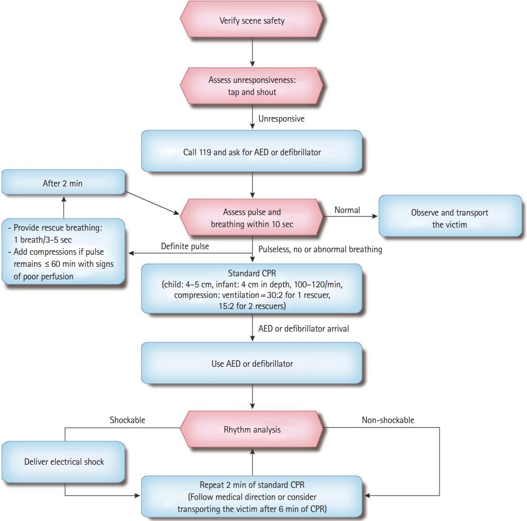
PBLS FOR HEALTHCARE PROVIDER
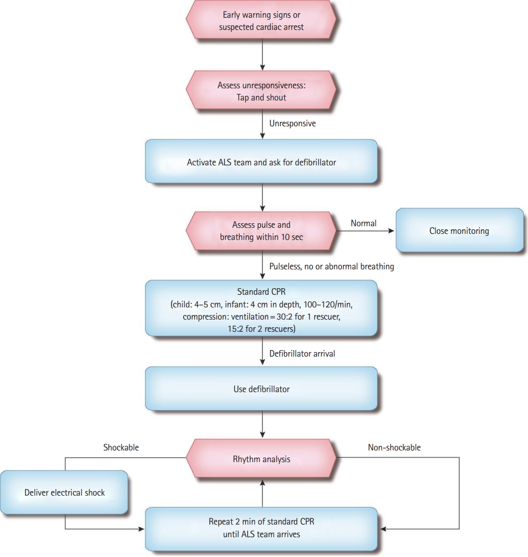
PBLS for healthcare provider has certain differences from for the lay rescuer; however, they are similar (Table 2 and Figs. 4, 5) [18,19]. Healthcare providers are mostly working in teams; therefore, each process is often simultaneously performed (chest compression and preparation of ventilation). Therefore, the priority of the activity is relatively less emphasized.
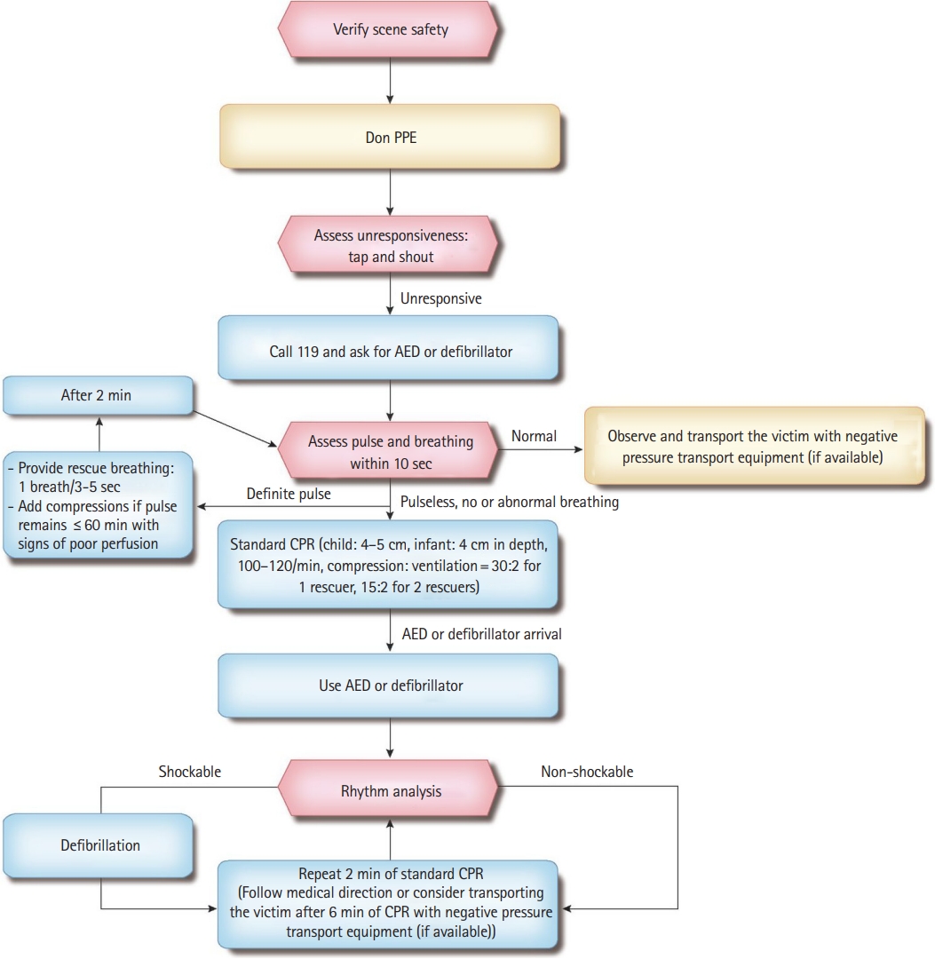
Safety of rescuer and patient
A rescuer should verify the safety in the current area whenever CPR is performed. Healthcare providers during the COVID-19 pandemic are recommended to use personal protective equipment (PPE) while performing aerosol-generating procedures in CPR. It is recommended that healthcare providers wear appropriate PPE, including masks, gloves, goggles, and surgical gowns, because chest compression and ventilation can cause the formation of aerosols and increase the risk of transmission of infection. When defibrillation is strongly recommended, it is suggested to actively implement it while paying attention to the transmission of infection (Fig. 6). Although compression-only CPR has a low risk of transmission of infection, it is suggested to cover the patient’s mouth if possible because there is a potential for aerosol generation. The rescuer should wash hands with soap and water or disinfect his hands with alcohol-based hand sanitizer as soon as possible as per infection control rules, and then change his clothes after finishing CPR. Moreover, the rescuer should contact local health authorities to verify for COVID-19 tests and self-isolation.
Check the response
First, verify whether the patient requires CPR. Tap the patient lightly and shout, “Are you okay, kid?” If you know the name of that kid, call his name. Quickly verify whether the child is injured and what medical treatment is required.
Activation of the EMS
If the patient is unresponsive and does not breathe (or gasping/abnormally breathing), ask someone nearby to call 119 and bring an AED. In the hospital, a witness immediately asks for help, ask a nearby person to bring a defibrillator, verify the pulse and breathing of the patient within 10 seconds, and start CPR if there is no pulse or breath.
Check the pulse
If the infant or child has no response and is not breathing normally, the care provider should check his pulse. The pulse check time should not exceed 10 seconds. The pulse is verified in the brachial artery in infants and the carotid or femoral artery in children. If you cannot feel the patient’s pulse within 10 seconds or are unclear if there is a pulse, start chest compression [20].
Insufficient breathing and adequate pulse with good perfusion
If the patient’s pulse is >60/min but breathing is inappropriate, provide rescue breathing at a rate of 12–20/min until spontaneous breathing is recovered (breathing once every 3 to 5 seconds). The pulse is then rechecked every 2 minutes and the pulse checking time should not exceed 10 seconds.
Bradycardia with poor perfusion
If the patient’s pulse is < 60/min and the perfusion is poor even with oxygen and ventilation (i.e., the skin is pale, mottled, or cyanotic), start chest compression. Slow pulse with poor perfusion is a sign of the requirement for chest compression in infants and children because cardiac output in them largely depends on the heart rate (HR). CPR should be immediately performed before the occurrence of cardiac arrest to improve survival rate. The absolute criterion for HR at which chest compression should be started is not yet clear. It is recommended to perform chest compression when HR is < 60/min and the perfusion is not good for the convenience of education and memory of skills.
Chest compression
If the infant or child is not responding and there is no breathing and pulse (or if it is unclear whether there is a pulse), start chest compression. The difference in method used by the healthcare provider and that by the lay rescuer lies in how to compress the chest in infants. When the healthcare provider is alone, two-finger chest compression is used for infants. When there are two or more rescuers, two-thumb encircling chest compression is performed. It is to stretch the hand and wrap the infant’s rib cage with both hands and strongly press the compression point of the sternum with both thumbs side-by-side.
The advantage of the two-thumb encircling chest compression is that it increases the coronary artery perfusion pressure more than that by the two-finger chest compression. It can consistently maintain adequate compression depth and force and can produce higher systolic and diastolic pressures. If you cannot hold the patient’s rib cage with both hands, just press the chest with two fingers.
The location of chest compression is the sternum right below the line connecting the nipples in infants and the lower half of the sternum in children. For one rescuer, the ratio of chest compression-to-ventilation is 30:2 using the two-finger chest compression. For two or more rescuers, the ratio of chest compression-to-ventilation is 15:2 using two-thumb encircling chest compression.
Airway opening and ventilation
Open the airway by tilting the head and lifting the jaw, and perform rescue breathing twice after 30 chest compressions (15 compressions if there are two rescuers). If there are signs of trauma that suggest spinal damage, open the airway with a jaw lift rather than a head tilt. It is very important to open the airway and perform ventilation in pediatric resuscitation; therefore, if you cannot open the patient’s airway with only jaw-lifting, you should apply the head tilting regardless of the trauma.
If it is difficult to breathe through the mouth and nose at the same time when giving ventilation to infants, mouth-to-mouth or mouth-to-nose ventilation can be performed. For mouth-to-mouth ventilation, make the patient breathe through the mouth while pinching the patient’s nose. Confirm whether the patient’s chest rises when inhaling from both sides.
Equipment and methods related to ventilation
Protective equipment
As some rescuers are reluctant about the direct contact by mouthto-mouth ventilation, they should use PPE. The use of PPE is not 100% effective in preventing the spread of infection and can create resistance to airflow. Therefore, ventilation should not be delayed in using PPE.
Bag-mask ventilation (BMV)
Most pediatric cardiac arrest patients can have adequate ventilation with bag mask breathing; however, it has the disadvantage of frequent interruption of chest compressions and a high risk of airway aspiration compared with the insertion of an advanced airway (endotracheal intubation or SGA). But, it requires more time to master the intubation or SGA skills than to master BMV. In pediatric OHCA, intubation failure rates and complications are higher than those in the BMV. From these points, it is reasonable to apply the BMV rather than intubation or SGA for rescue breathing of pediatric OHCA patients. It is important to learn techniques such as selecting the appropriate size for the mask, opening the airway, fitting the mask close to the face, and applying appropriate pressure for effective BMV.
Ventilation bag
Pediatric self-inflating bags supply at least 450 to 500 mL air, and the bags with a smaller capacity may not provide sufficient tidal volume for term infants. For older children or adolescents, an adult self-inflating bag (approximately 1,000 mL) should be used. If oxygen is not supplied, it is ventilated only with room air; if the amount of oxygen supplied is 10 L/min, the oxygen concentration is maintained from 30% to 80%. To supply a higher concentration (60%–95%) of oxygen, connect an oxygen reservoir to the bag. Oxygen at 10–15 L/min can be supplied to the reservoir attached to the pediatric bag and at least 15 L/min to the adult bag.
Ventilation method to prevent hyperventilation
Hyperventilation reduces circulating blood flow; therefore, it is important to prevent it. Two ventilations are performed after 30 chest compressions (by one rescuer) or 15 chest compressions (by two healthcare providers) before using advanced airway (insertion of an endotracheal tube or SGA devices is achieved); at this time, mouth to mouth ventilation or BMV is used. After performing advanced airway management, the “compression to ventilation ratio” of CPR is not used. Continue chest compressions at a rate of 100–120/min without interruption and ventilation 10 times per minute. Two or more healthcare providers should rotate the compression role every 2 minutes to prevent exhaustion. If the patient has ROSC but has no breathing, perform only ventilation 12–20 times/min (once per 3–5 seconds). Excessive ventilation during CPR decreases venous return, thereby reducing cardiac output and cerebral blood flow and reducing coronary artery perfusion because of an increase in intrathoracic pressure. Therefore, the rescuer must ventilate according to the number per minute. Hand-compressed bags can generate high pressure; therefore, ventilate only enough to observe the chest rise.
Two rescuers BMV
BMV together by two rescuers helps with effective breathing when there is severe airway obstruction and poor lung elasticity or when it is difficult to fit the mask to the face tightly. One rescuer maintains the airway open with both hands and attaches the mask firmly to the patient’s face and the other rescuer squeezes the ventilation bag. Two rescuers should verify whether the patient’s chest is rising.
Gastric distention and applying cricoid pressure
You should avoid gastric distention because it impairs efficient ventilation and may cause vomiting. To minimize gastric distension, every breath can be performed over a second to avoid overpressure during inhalation.
Applying cricoid pressure may be considered to prevent air from entering the stomach. However, it is not recommended to routinely perform cricoid pressure. Cricoid pressure should be considered only when the patient is unconscious and there is another healthcare provider. Moreover, be aware that excessive pressure on the cricoid cartilage can block the trachea.
Oxygen
Animal experiments show that 100% oxygen is harmful, but human studies show no detrimental effects of oxygen concentration after the neonatal period; therefore, supply 100% oxygen during resuscitation. When the patient is stabilized, supply oxygen while verifying the oxygen concentration. The administration of humidified oxygen can prevent dryness of the mucous membranes and thickening of lung secretions.
Oxygen is administered using a mask or nasal cannula depending on the patient’s breathing condition. The mask supplies 30% to 50% oxygen when spontaneous breathing occurs. High concentrations of oxygen can be administered by supplying 15 L/min of oxygen with a mask that tightly fits on the face and has a reservoir. When there is spontaneous breathing, use a nasal cannula suitable for the size of infants and children. When using a nasal cannula, up to 0.5–1 L/min can be administered for neonates, 1–2 L/min for infants, 4 L/min for preschool children, and 6 L/min for school-age children. The oxygen concentration is adjusted as per the child’s size, respiratory rate, and breathing effort. When 2 L/min of oxygen is administered to an infant, the inhaled oxygen concentration is 55% [21].
Defibrillation
Ventricular fibrillation (VF) and pulseless ventricular tachycardia (pVT) may be the cause of sudden cardiac arrest or occur during CPR. A sudden collapse in children with witnesses (e.g., collapse during exercise) is attributed to VF or pVT, requiring immediate CPR and rapid defibrillation. VF and pVF are classified as “shockable rhythm” because they respond to defibrillation.
For infants, it is beneficial to use a manual defibrillator if a well-trained healthcare provider has identified the shockable rhythm. The first energy dose is 2 J/kg and the second one is ≥4 J/kg during defibrillation, which should not exceed 10 J/kg or the maximum dose for adults [22].
If you do not have a manual defibrillator, use an AED with a pediatric impulse attenuator for infants. Use an AED with a pediatric energy attenuator or pediatric patches for children aged <8 years. However, if you have neither a manual defibrillator nor an AED with an energy attenuator, an AED for adults that cannot control the dose of energy can be used for infants.
Rescuers should minimize the interval time between chest compressions and defibrillation and resume CPR by immediately restarting chest compressions after one defibrillation. The AED encourages the rescuer to reanalyze the rhythm every 2 minutes, and the defibrillation should ideally be immediately performed after the chest compression.
RESUSCITATION IN SPECIAL CIRCUMSTANCES
Airway obstruction (choking) by foreign bodies
Note that >90% of deaths by a foreign body aspiration occur in children aged <5 years, of which 65% occur in infants [23]. The clinical symptoms of airway obstruction by a foreign body include sudden shortness of breath, coughing, nausea, grunting, and wheezing. It suddenly occurs and has no previous fever or respiratory symptoms, which distinguishes it from other respiratory difficulties. Airway obstruction caused by foreign bodies can range from mild to severe. If airway obstruction is mild, the child may cough or make a sound. Symptoms suspected of complete obstruction include no speech, no sound when coughing, unable to breathe, cyanosis, or loss of consciousness; in this case, immediate treatment is required.
If it is determined that airway obstruction is severe in children aged >1 year, perform back blow until a foreign body comes out or consciousness disappears [24]. For infants, perform the back blow five times and chest thrust five times until a foreign body comes out or the consciousness disappears repeatedly [25,26]. Abdominal thrust is not performed in infants owing to the high risk of internal organ damage because the ribs do not sufficiently protect the epigastric organs and the liver is relatively large [25].
If the patient does not respond or his reaction disappears during the foreign body removal procedure, start CPR regardless of the presence or absence of a pulse [27]. However, if you can see a foreign body in the patient’s mouth before breathing after chest compression, use your fingers to remove it. If you cannot see a foreign body in the patient’s mouth, do not try to remove it using your fingers. This blind swabbing of the mouth may push the foreign body deeper into the pharynx or damage the pharynx [27,28]. After two ventilations, perform chest compression and ventilation repeatedly until the foreign body is removed. In fact, there are limited high-quality studies that can help determine guidelines for airway obstruction by foreign bodies. Most airway obstruction caused by a foreign body is resolved by letting the patient cough himself. However, in severe cases, the patient may need help from a rescuer [29,30].
Trauma
Unintended trauma can be a leading cause of death in children and adolescents. Cardiac arrest by major blunt and penetrating injuries in children has a very high mortality rate [31-33]. Tension pneumothorax, hemothorax, lung injury, and cardiac tamponade can interfere with hemodynamics, oxygenation, and ventilation. Therefore, you should always suspect severe chest injury in patients with chest-abdominal trauma. Early treatment for reversible causes after cardiac arrest owing to penetrating trauma can increase survival [34,35]. It is recommended in the guidelines to control bleeding, recover circulating blood volume, secure airways, and treat tension pneumothorax for cardiac arrest caused by trauma. These procedures should be concurrently performed with standard CPR. The principles of basic CPR for injured children are the same as for children with common diseases; however, there is certain emphasis. Improperly performed CPR can increase preventable mortality. Errors in opening and maintaining the airways and errors caused by not recognizing internal bleeding are common in resuscitating children. The points to be noted when performing CPR in pediatric trauma patients are listed below [36]. 1) Cardiac arrest in pediatric trauma patients often occurs because breathing is not adequately maintained owing to hypovolemia by trauma. Therefore, it must be determined whether the airway and ventilation are maintained even in trauma patients. 2) If there is a possibility of airway obstruction because of broken teeth or blood, use a suction device. 3) If there is external bleeding, press to stop it. You should remove the patient’s clothes and check his entire body to determine the bleeding points. After verification, ensure to cover with a warm cloth such that hypothermia does not occur. 4) If there is a possibility that a spinal injury occurred judging by the mechanism of the injury, then minimize cervical spine movement and do not pull or move the head and neck. Open the airway by lifting the patient’s jaw and do not tilt the head. If the airway is not maintained by lifting jaw, secure the airway by tilting the head and lifting the chin. If there are two rescuers, one opens the airway and the other prevents cervical spine movement. At least the thighs, pelvis, and shoulders are fixed together on the spinal board. Because infants and toddlers have relatively large heads, the occipital part should be placed in a slightly recessed position than the torso, or the torso should be laid in a slightly elevated position and fixed to the spinal correction board to prevent cervical flexure [37]. 5) Transfer children with multiple organ trauma to a trauma center with a pediatric specialist if possible. 6) Open thoracotomy could be considered for children with no pulse with a penetrating injury [2,34,38-40]. However, there is still insufficient evidence to recommend performing emergency thoracotomy in children and infants without a pulse because of blunt trauma [41,42].
Drowning
Drowning time is an important predictor of prognosis. Age, promptness of first aid, water type (freshwater or seawater), water temperature, and presence of witnesses will not be reliable prognostic factors [42]. For drowning in ice water, the rescue time can be extended because there is a possibility of survival even if the drowning time is long [43,44]. After removing the drowned children from water, initiate CPR immediately. Rescuers with special training initiate rescue breathing while underwater. Do not perform chest compressions in water because they are ineffective [45].
There is no evidence that water acts as a foreign body causing airway obstruction. Therefore, you do not waste time trying to drain water from the patient’s lungs [46]. Open the airway, perform rescue breathing twice, and start CPR with chest compressions. If there is one rescuer, perform chest compressions and ventilations five times cyclically at the ratio of 30:2, call the EMS, and ask for an AED. If there are two rescuers, the first rescuer continues CPR, whereas the second rescuer reports to the EMS and prepares an AED [47].
Children who require special medical assistance
Children who require special medical assistance because of complications owing to chronic disease (e.g., blockage of tracheostomy), problems with auxiliary medical devices (e.g., ventilator malfunction), and exacerbation of the existing disease need appropriate management for their conditions. If there is no information on an existing disease, treatment plans, and current medications, you may have difficulty performing treatments. Parents or caregivers should copy the child’s medical information and place it at home, school, or care facility in advance. If the child is discharged from the hospital with a chronic or fatal illness, parent, school nurse, and home care provider should be aware of information about the reason for hospitalization, the condition during the hospitalization, and any symptoms that may worsen as well as receive training for CPR in special circumstances [48].
Children with prior medical directions
If a decision has been made to limit or abandon CPR, medical personnel should specify CPR-related limitations in detailed prescribing instructions. The doctor should separately specify the prescribing instructions for situations outside the hospital. Care facilities and school nurses should copy and place the form of a child for whom decision of giving up on resuscitation attempts has been made. Parents, school nurses, and home-visit health care providers should receive sufficient information about the guide of CPR and the contact information in case of an emergency [49].
Ventilation through tracheostomy or tracheostomy window
Caregivers of a child who received a tracheotomy (parent, school nurse, and home health care provider) should know how to maintain the child’s airways, how to remove airway secretions, and how to perform CPR through artificial airways. They should perform assisted ventilation through a tracheostomy window and verify the airway maintenance of the child and whether his chest is rising. If ventilation through the tracheostomy window is not effective even with suction, it should be suspected that the tracheostomy tube was not properly inserted and airway maintenance should be checked again. When the patient’s breathing is not appropriate, perform mouth to tracheostomy window ventilation for the patient who has had a tracheostomy. If the tracheostomy window is already blocked, perform BMV via the nose and mouth [50].
PREVENTION OF PEDIATRIC CARDIAC ARREST
Rapid response system for preventing pediatric cardiac arrest
Rapid response systems have been employed in several medical institutions to prevent cardiac arrest by detecting physiological changes in hospitalized patients early and to reduce admission to the intensive care unit [51]. This is to identify the clinical deterioration of the patient due to a delay of a call to medical staff in a hospital. It is an active effort to issue an early warning of changes in a patient’s condition by scoring blood pressure, pulse, respiratory rate, body temperature, oxygen saturation, and level of consciousness [52].
The use of a pediatric early warning score has reduced the incidence of cardiac arrest, length of stay in the hospital, and mortality as well as unplanned transfer to the pediatric intensive care unit [53,54]. However, there are not many studies on the frequency of early response as per the working hours of the rapid response team (RRT) and the patient’s prognosis, and there is currently no consensus on the standard for activating the domestic RRT; therefore, continuous further study is required [55].
Moreover, when using pediatric early warning scores in medical institutions, it is necessary to discuss how to organize items to sensitively identify changes in the patient’s condition. It is necessary to discuss whether to include only vital signs or also include sepsis, dyspnea, anaphylactic shock, hypovolemic shock, obstructive shock, arrhythmia, and metabolic acidosis [48]. Moreover, it is necessary for the medical staff to quickly recognize and respond after discussing how to identify the false positive alerts of early warning score.
Pediatric RRT (or medical emergency team)
RRT is a department dedicated to first aid and monitoring of patients with deteriorating conditions in the hospital. It is composed of specialized medical staff to quickly identify and respond to signs that appear before a cardiac arrest occurs. Depending on the hospital’s resources, the composition, operation method, and activation of the rapid response system can be adpated. Medical institutions form RRTs to better understand the patient’s respiratory system, cardiovascular system, and changes in consciousness, and it comprises skilled medical staff for rapid intervention and efficient critical care management [56,57].
There is not sufficient evidence that the operation of the pediatric RRT increases the rate of ROSC and decreases the mortality rate of pediatric cardiac arrest in hospitals. However, studies are showing that it reduces the incidence of pediatric cardiac arrest in hospitals, particularly outside the intensive care unit [58-61]. In one cohort study, for children requiring unplanned admission to the pediatric intensive care unit, RRT implementation was associated with reduced mortality [62]. Therefore, medical institutions with trained professional staff may consider establishing and operating a pediatric RRT to prevent the occurrence of cardiac arrest.