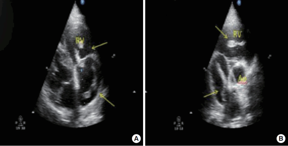
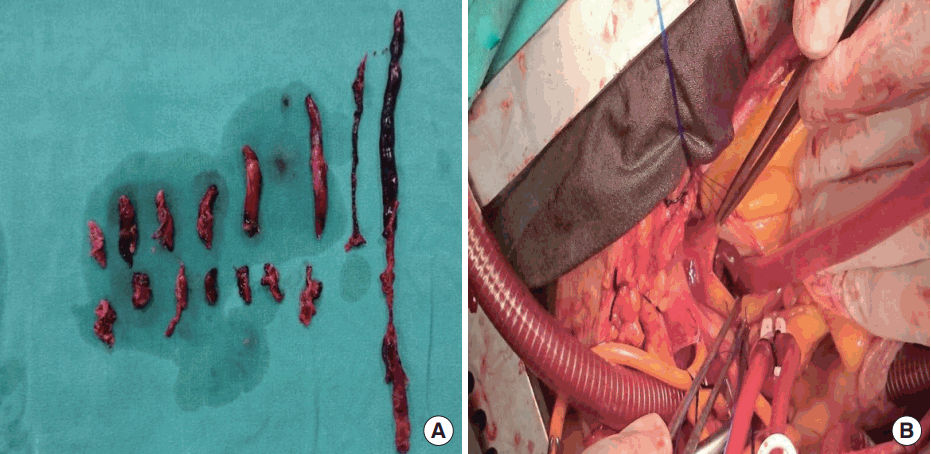
A 51-year-old man presented to the emergency department with progressive dyspnea and orthopnea that had persisted for 15 days. The patient had no history of any disease. Upon physical examination, he had a brachial blood pressure of 80/50 mmHg, a heart rate of 130/min, and a respiratory rate of 40/min; a 2/6 pansystolic murmur was heard in the mesocardiac region, and breath sounds were decreased in the patientŌĆÖs lungs. An electrocardiography revealed sinus tachycardia and a right bundle branch block. The patientŌĆÖs arterial blood gas measurements were as follows: pH, 7.52; pO2, 61.8 mmHg; pCO2, 20.5 mmHg; HCO3-, 16.6 mmHg; and O2 saturation, 93%. With regards to other blood parameters, biochemical analysis revealed a hemoglobin level of 11.4 g/dL, a white blood cell count of 12,990 /╬╝L, a platelet count of 169,000 /╬╝L, a serum creatinine level of 0.97 mg/dL, a creatinine kinase-MB level of 2.78 ng/mL (within the normal range), a troponin I level of 0.449 ng/mL, and a D-dimer level of >5,000 ng/mL. We carried out echocardiography, which revealed a 60% left ventricular ejection fraction, a moderated tricuspid regurgitation, and a systolic pulmonary artery pressure of 70 mmHg, with an enlarged right ventricle. The interventricular septum was semi-paradoxical, and the right ventricular free wall was hypokinetic. A 7├Ś1.5 cm, free-floating giant thrombus was seen extending from the inferior vena cava to the right atrium and right ventricle (Fig. 1). A mobile thrombus was also seen in the proximal right pulmonary artery. We diagnosed acute, high-risk pulmonary embolism. The patient underwent emergency surgical thrombectomy, and the thrombus was removed (Fig. 2).
Pulmonary embolism (PE) is a common, potentially fatal condition. Early diagnosis and anticoagulation therapy can reduce mortality from 30% to 2% to 10% [1,2]. However, in emergency rooms, it is not always possible to access the definitive diagnostic tools for acute pulmonary embolism. Arterial blood gas analysis, electrocardiogram, and chest radiography are not reliable tools for detecting PE. Moreover, D-dimer testing is less useful in patients who are already hospitalized, because D-dimer levels are elevated in many illnesses that mimic PE [3]. Scintigraphy and multidetector computed tomography might be difficult and costly to perform and interpret in emergency departments in some countries. In addition, they are not suitable for every patient (particularly for unstable patients); contrast agents is another problem with these techniques.
Transthoracic echocardiography is a useful method for diagnosing PE in emergency rooms. It can be used to evaluate right ventricular morphology and function, as well as paradoxical movement of the interventricular septum. It is a rapid, practical, and sensitive technique for quickly identifying right heart thrombi. Echocardiography may also help to exclude other diagnoses, such as aortic dissection, pericardial disease, myocardial infarction, and valvular insufficiency [4]. In unstable patients, emergency department doctors can perform the technique quickly at the bedside.
Giant, mobile, thrombus-related pulmonary embolisms are rare. If the giant thrombus is in the right atrium, mortality can be as high as 45% [5,6]. Indeed, the mortality rate is 80% to 100% in untreated patients with mobile thrombus in the right side of the heart. The treatment options for these patients include fibrinolytic therapy, surgery, and anticoagulant therapy [7]. If an emergency physician diagnoses PE quickly using ultrasound, he/she will be able to treat the patient quickly, especially if surgery is necessary.