INTRODUCTION
Catheter tip malpositioning into other central veins may increase the risk of chemical or bacterial venous thrombophlebitis and vascular perforation [1]. In addition, accurate central venous pressure monitoring will be impossible [2-4]. When a subclavian central venous (SCV) catheter is misplaced into the ipsilateral internal jugular vein (IJV), catastrophic events such as encephalopathy or infection may occur due to elevated intracranial pressure or thrombosis associated with the misplacement [4-7].
Compared to the IJV route, the SCV route has a greater chance of misplacement [8-10]. To decrease the occurrence rates of SCV catheter misplacement, using the Ambesh maneuver to orient the needle bevel and shoulder and head positions are considered effective to a certain degree [2,11,12]. Still, the reported incidence of unintentional positioning of a central venous catheter tip in other central veins ranges from 2% to 21% [11-14]. Confirmation of the misplacement could be made using bedside techniques such as saline flush testing, pressure monitoring, intravascular electrocardiography, and ultrasonographic confirmation [1,15-18]. Use of these methods is largely limited as they are not routine methods in emergencies; therefore, it is important to prevent catheter misplacement.
Previous studies have reported comparisons between misplacement rates and patientsŌĆÖ shoulder positions [19-21]. A recent randomized controlled trial suggested that the neutral position was associated with a reduced misplacement rate, but the preferred shoulder position (lowered, elevated, or neutral) for misplacement prevention remains controversial [20].
To date, there has been no quantitative evaluation of shoulder position in the context of catheter misplacement. We attempted to evaluate geometric variables such as the clavicular tilt angle (CTA) obtained from anteroposterior (AP) chest X-ray images to determine their correlations with patientsŌĆÖ shoulder positions and investigate the relation of such variables with the chance of catheter misplacement. Although limited, our study introduced a quantitative method for and insight into investigating SCV catheter misplacement, a potential problem plaguing emergency physicians [22,23]. The present study was conducted to investigate the relation between shoulder position and SCV catheter misplacement. Shoulder position was estimated using CTA values observed on AP chest X-ray images.
METHODS
Study design and setting
A retrospective case-control study was conducted at a 1,080-bed academic tertiary referral center. The annual emergency department (ED) volume is 90,000 visits, including 70% adult visits. The hospital has been using electronic medical records and picture archiving and communication system (PACS). The present study was approved by the hospitalŌĆÖs Institutional Review Board, and informed consent was waived.
Selection of participants
The study was conducted on all the adult patients (>18 years of age) who underwent central venous catheterization in the ED of our hospital between January and December 2012. We closely investigated cases of SCV catheter misplacement. Catheterization was performed using double/triple lumen non-tunneled infusion catheters (ARROWguard, Blue PLUS central venous catheter, Arrow International Inc., Reading, PA, USA). Exclusion criteria were (1) patients who underwent catheterization via the Ambesh maneuver; (2) those who had tunneled dialysis catheters; and (3) those who had rib or clavicle fractures.
All central venous catheterizations were conducted using a modified Seldinger technique without ultrasonographic guidance at the patientsŌĆÖ bedside. The catheterization procedure applied during the study period was as follows: (1) Place the patient in a supine position and the patientŌĆÖs head in a neutral position. (2) Clean the neck and infraclavicular areas including the suprasternal notch with 0.5% chlorhexidine solution. Isolate them with sterile drapes. (3) Anesthetize the region by applying 1% lidocaine around the midpoint of the clavicle. (4) Place the index finger and the thumb of the non-dominant hand on the suprasternal notch and the medial two-thirds of the clavicle, respectively. Find the puncture point directly below, where the lateral third of the clavicle meets the medial two-thirds. (5) Direct the introducer needle with the bevel facing down towards the suprasternal notch at a 10┬░ angle to the surface of the chest. (6) Advance the 18 G introducer needle slowly through the skin and subcutaneous tissues until flush blood or dark venous blood appears. Seek help from a more experienced physician after three failed attempts. (7) Check for continual free venous flow with aspiration. (8) Insert a J-tip guidewire while maintaining a caudal direction and remove the introducer needle. (9) Dilate the skin and subcutaneous tissues overlying the guidewire using a dilator. (10) Railroad a central catheter over the guidewire 12ŌĆō14 cm into the subclavian vein. (11) Remove the guidewire. (12) Confirm catheter functions and suture the catheter. (13) Apply a sterile transparent dressing. (14) Confirm correct catheter placement on a chest X-ray image.
In our study, experienced physicians were those who performed the above catheterization procedure 50 or more times [24,25]. For every patient with a misplaced catheter, two other patients who underwent catheterization within 30 days of the time when the misplacement occurred were matched as the control group under two conditions: (1) they were the same sex as the misplacement case, and (2) the difference in ages was minimum.
Data collection and processing
Demographic, clinical, and radiological data were extracted from patient medical records and the AP chest X-ray image database in a standardized fashion. We performed a 1:2 matched case-control study to constrain possible confounding factors. We randomly selected 20% of the records for an independent review by an abstractor. Discrepancies arising from the review of the results by other data abstractors were resolved with the help of a radiologist or a senior emergency physician. The collected data included age, sex, diagnosis, catheterization side, physicianŌĆÖs level of experience, and the occurrence of catheter misplacement. Catheter misplacement was confirmed on AP chest X-ray images by examining the catheter tip. The intended position was between the SCV and the right atrium. Malpositioning was typically found in the ipsilateral IJV, contralateral brachiocephalic vein (BCV), or contralateral IJV.
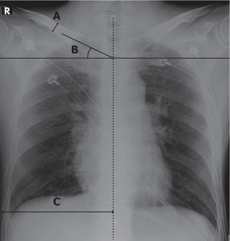
Geometric variables were estimated from the X-ray images, and they included the ipsilateral transverse length of the thorax (ITL-T), CTA, and thickness of the clavicle (TC). TC was defined as the superior-inferior width at the midpoint between the sternal end and acromial end of the clavicle. The CTA was defined as the angle between two vectors, i.e., the line bisecting the proximal portion of the clavicle and the horizontal line [22,24]. CTA is a geometric variable for evaluating shoulder balance/imbalance [22,23]. The ITL-T was defined as the longest transverse length between the spinous process of the thoracic vertebrae and the most laterally located rib shadow. In Fig. 1, these variables are displayed on a chest X-ray image. Geometric measurement values were entered into the software analysis as tabulation (Excel 2013, Microsoft Co., Redmond, WA, USA).
Primary data analysis
Inter-rater reliability was assessed using the generalized ╬║-statistic and 95% confidence interval (CI) for four data elements: catheter misplacement and appropriateness of CTA, TC, and ITL-T. All data were expressed as mean┬▒SD as appropriate, and they were analyzed using a statistical tool (Stata IC ver. 11, Stata Co., College Station, TX, USA). The significance of inter-group differences was assessed with the chi-square test for categorical variables and the t-test for continuous variables. A two-sided P-value less than 0.05 was considered to indicate statistical significance.
RESULTS
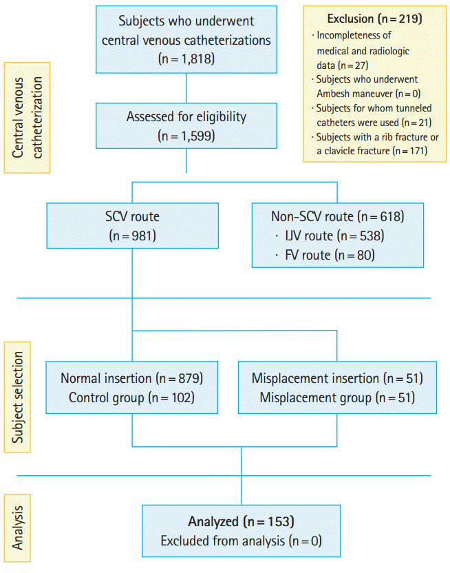
A total of 1,818 patients underwent central venous catheterization during our study period, and 1,599 cases were considered for inclusion (Fig. 2). Of these catheterizations, the subclavian route was used 981 times (61.4%) and misplacement occurred 51 times (5.2%). Misplacement into the ipsilateral IJV occurred 43 times (right, 41; left, 2) and misplacement into the contralateral BCV occurred 8 times (right, 8; left, 0). No misplacement into the contralateral IJV occurred. Sixteen of the misplacements were by inexperienced physicians (less than 50 performances) and 35 were by experienced physicians (more than 50 performances).
Table 1 shows patient demographics and AP chest X-ray evaluation results. TC values in the misplacement group and the control group were 13.2┬▒2.1 mm and 13.7┬▒2.8 mm, respectively (95% CI, ŌĆō1.38 to 0.35; P=0.20). CTA values in the misplacement group and the control group were 28.5┬░┬▒7.3┬░ and 22.6┬░┬▒6.3┬░, respectively. The CTA value in the misplacement group was significantly greater than that in the control group (95% CI, 3.63 to 8.11; P<0.001). ITL-T values in the misplacement group and the control group were 137.0┬▒11.6 mm and 138.7┬▒13.7 mm, respectively (95% CI, ŌĆō6.12 to 2.71; P=0.45).
The four data abstractors in our study reached agreement for more than 95% of the data elements. The value of Cohen ╬║- was 0.99 for catheter misplacement (95% CI, 0.96 to 1.00), 0.80 for TC (95% CI, 0.67 to 0.94), 0.89 for CTA (95% CI, 0.72 to 0.95), and 0.94 for ITL-T (95% CI, 0.77 to 1.0).
DISCUSSION
Catheter insertion through the SCV is frequently performed in the ED, and catheter tip malpositioning should be avoided for improving patient prognosis. Previous studies of SCV catheter insertion mostly focused on factors associated with successful puncture of the SCV, catheterization failure, and positions of the malpositioned catheter tips along with the associated complications [1,4,8,19,24,25]. In the present study, we attempted to associate incidences of misplacement with radiographic features of patient shoulders. The incidence of catheter misplacement in our study was 5.2%, which falls in the range of previously reported values: 21.4% by Tripathi et al.[12], 5.4% by Sanchez et al. [13], and 2.1% by Ambesh et al. [14]. Inconsistencies of these percentage values must be associated with various confounding factors such as the direction of the J-tip and varying competency levels of the performers, to name a few. In our study, all catheterizations strictly followed the standard protocol described above. Heretofore, the methods suggested for successful SCV catheterization included caudal positioning of the guidewire tip, caudal positioning of the plunger end after puncture, Ambesh maneuver, head positioning, and inferior positioning of the shoulder [12-14,26,27]. The Ambesh maneuver refers to a manual compression of the ipsilateral IJV at the time of guidewire insertion to prevent its progression into the ipsilateral IJV [11]. However, these methods are not always successful.
A quantitative study of shoulder position based on computed tomography images in the context of catheter misplacement was performed by Kitagawa et al. [19]. They found that a superior-inferior change of 5 cm in shoulder height increased the overlapping area between the clavicle and SCV from 33.5% to 40%, increased the angle between the SCV and IJV from 89┬░ to 117┬░, and decreased the angle between the SCV and BCV from 114┬░ to 99┬░ in the neutral shoulder position. These results imply that the shoulder should be kept in the inferior position, which will lead to a maximal overlapping region between the clavicle and the SCV, and a successful puncture of the SCV [19]. However, controversy regarding this issue remains since Baltalarli et al. [21] reported that maintaining a superior shoulder position resulted in higher success rates of SCV catheterization, although a relatively more consistent relationship between the clavicle and the SCV could have been a confounder.
We noted a lack of studies that approached this problem quantitatively. In this study, we measured CTA values from AP chest X-ray images as described in Fig. 1A, and a CTA increase increase is likely to be associated with an upward change of shoulder position, while a CTA decrease is likely associated with a downward change. Mean CTA measurements were 28.5┬░ and 22.6┬░ in the misplacement and control groups, respectively. The CTA in the misplacement group was 5.9┬░ greater than that in the control group, and the difference was significant. This result suggests that decreasing the CTA and maintaining an inferior shoulder position could be important for reducing the risk of misplacement in SCV catheterization. It seems that the CTA is determined not only by shoulder position, but potentially also by other factors such as ITL-T, AP length of thorax, clavicle length, height, and body weight. Thus, future studies should evaluate the effect of the AP length and the length of the clavicle on SCV catheterization.
This retrospective study has many limitations. First, different levels of procedure competency may have affected the incidence rates of misplacement. This may not have been a major confounding factor, however, as the occurrence of misplacement in the present study falls within a reasonable range (2% to 21.4%, as previously reported) [12-14]. In addition, in our hospital, all procedures strictly followed the standard protocol. Second, the chest X-ray images were obtained at a follow-up after the catheter insertion. Therefore, the CTA values may not precisely match the actual shoulder angles at the time of insertion. This may have played a limited role as a confounder, as the same conditions were imposed on both groups. Third, possible confounders include respiratory movements, the physical condition of patients at acute clinical stages, and unadjusted directional characteristics between AP and PA. Fourth, our study is largely limited by the fact that only a univariate analysis was included in the study design. Future studies should address these issues.
Positional characteristics of the clavicle and the scapula are thought to be major factors determining patient shoulder position [22,23,28]. In our study, CTA alone was found to be associated with a higher occurrence rate of misplacement. Although limited, our investigation incorporated the use of geometric measures based on X-ray images such as CTA values. We believe that such variables provide ED clinicians with a useful quantitative insight into the issue of catheter misplacement.
In summary, radiological evaluation found that the CTA was larger in the misplacement group than in the control group. Assuming that CTA indicates the actual shoulder position, our findings suggest that SCV catheter misplacement should occur less frequently by avoiding patient shoulder elevation.